![]()
Built for Efficiency
The Avanti system is designed to scan patients faster with 70,000 A-scans per second, rapidly process scans with Avanti’s background processing engine, and quickly identify potential problems with comprehensive reports.
New Views of the Retina
Avanti Widefield OCT gives us information on retinal structures outside a traditional 6×6 mm cube, separates the retina into distinct layers for detailed assessment, offers views of the vitreous and deep choroid, and gives us the ability to monitor change over time. With the extensive information delivered by the Avanti System, we can tailor our approach to treatment and truly personalize patient care.
Extensive Nerve Fiber Layer Analysis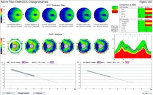
Avanti’s trend analysis software, which tracks change in RNFL and GCC thickness provides an estimate of future progression. This comprehensive analysis allows us to personalize treatment protocols and enhance your understanding of your disease. New metrics, Focal Loss Volume (FLV%) and Global Loss Volume (GLV%), increase the sensitivity and specificity of the GCC analysis to help us identify suspected optic nerve head disease.
Cornea Advance: Intelligent OCT Imaging for the Anterior Segment
Our OCT is expanded to address a broad range of patients in our practice. With Avanti’s Cornea Advance, we can visualize and measure corneal angles, quantify corneal thickness and track change in thickness between visits.
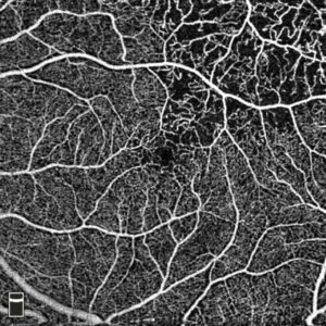 OCT Angiography: See the Retinal Vasculature
OCTA produces ultra-high resolution, three-dimensional images that are displayed as individual layers of retinal vasculature, allowing us to isolate specific areas of interest and see microvasculature that is not easily seen with traditional imaging methods. High-density OCTA imaging produces larger format scans with outstanding image quality to enable assessment with a wider field of view.
Measure Retinal and Optic Disc Vasculature to Enhance Patient Management
AngioAnalytics™ allows us to measure foveal avascular zone (FAZ) parameters, areas of abnormal flow in the outer retina and choroid, and vessel density of the superficial vascular complex, deep vascular complex and radial peripapillary capillaries. Multi scan analysis enables assessment of visit-to visit change, and trend reports predict change over time to bring powerful new information to our daily clinical practice.